Fil:HIV-budding.jpg
Hopp til navigering
Hopp til søk
Opprinnelig fil (2 967 × 1 971 piksler, filstørrelse: 717 KB, MIME-type: image/jpeg)
Beskrivelse
| Beskrivelse |
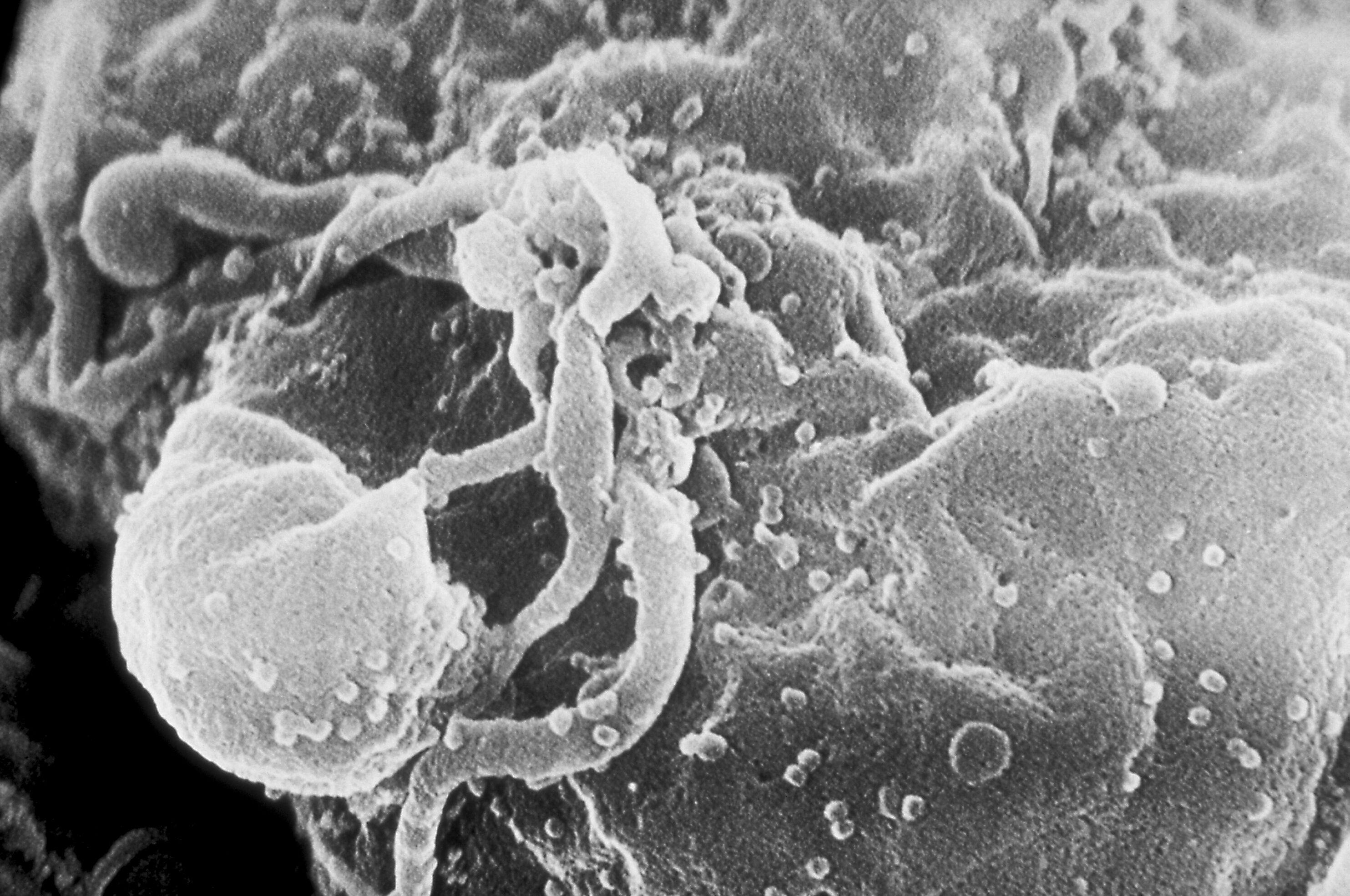
English: Scanning electron micrograph of HIV-1 budding from cultured lymphocyte.
Multiple round bumps on cell surface represent sites of assembly and budding of virions.
العربية: صورة بالمجهر الالكتروني لفيروس نقص المناعة البشري المكتسب متبرعم من خلية ليمفاوية Español: Viriones del VIH-1 ensamblándose en la superficie de un linfocito
日本語: リンパ球に結合するHIV-1
Polski: Wirus HIV pączkujący z hodowanego limfocytu. Obraz z elektronowego mikroskopu skaningowego
Italiano: Virus HIV in fase di gemmazione da una cellula umana
中文:掃描式電子顯微鏡視野下可見,HIV-1病毒正從培養出來的淋巴球出芽,準備進一步散佈開來
Suomi: Uusi HI-virus tulossa infektoituneesta isäntäsolusta
Magyar: Pásztázó elektronmikroszkópos felvétel tenyésztett limfocita sejten lévő HIV-1 vírusról
Bahasa Indonesia: HIV yang baru memperbanyak diri tampak bermunculan sebagai bulatan-bulatan kecil pada permukaan limfosit setelah menyerang sel tersebut; dilihat dengan mikroskop elektron
עברית: נגיף HIV מיקרוסקופ אלקטרונים סורק
Српски / srpski: Снимак вируса ХИВ електронским микроскопом
فارسی: اچ آی وی در حین جوانه زدن از یک گویچه سفید خون
Srpskohrvatski / српскохрватски: Snimak virusa HIV elektronskim mikroskopom
Deutsch: Zahlreiche HI-Viren, die aus einem kultivierten Lymphozyten knospen, SEM
Afrikaans: 'n Elektronmikrograaf van HIV-1 wat besig is om vanuit 'n gekultiveerde limfosiet te bloei
മലയാളം: എച്ച്.ഐ.വി. 1 വൈറസ് വികാസം പ്രാപിക്കുന്ന ചിത്രം |
||
| Dato | |||
| Kilde |
|
||
| Opphavsperson |
|
||
| Tillatelse (Gjenbruk av denne filen) |
PD-USGov-HHS-CDC English: None - This image is in the public domain and thus free of any copyright restrictions. As a matter of courtesy we request that the content provider be credited and notified in any public or private usage of this image. |
||
| Andre versjoner |
|


Orginal opplastningslogg
The original description page was here. All following user names refer to en.wikipedia.
- 2005-07-13 20:42 Carl Henderson (Corrected copyright tag.)
- 2005-07-13 20:40 Carl Henderson ('''Cite:''' Centers for Disease Control / C. Goldsmith '''URL:''' [http://phil.cdc.gov/PHIL_Images/1197/1197.tif http://phil.cdc.gov/PHIL_Images/1197/1197.tif] '''Description:''' Scanning electron micrograph of HIV-1 budding from cultured lymphocyte. Mu)
Lisensiering
This file is a work of the Centers for Disease Control and Prevention, part of the United States Department of Health and Human Services, taken or made as part of an employee's official duties. As a work of the U.S. federal government, the file is in the public domain.
|
Filhistorikk
Klikk på et tidspunkt for å vise filen slik den var på det tidspunktet.
| Dato/klokkeslett | Miniatyrbilde | Dimensjoner | Bruker | Kommentar | |
|---|---|---|---|---|---|
| nåværende | 14. apr. 2006 kl. 22:00 | Intet miniatyrbilde | 2 967 × 1 971 (717 KB) | wikimediacommons>Nikita~commonswiki | '''Cite:''' Centers for Disease Control / C. Goldsmith '''URL:''' [http://phil.cdc.gov/PHIL_Images/1197/1197.tif http://phil.cdc.gov/PHIL_Images/1197/1197.tif] '''Description:''' Scanning electron micrograph of HIV-1 budding from cultured lymphocyte. Mu |
Filbruk
Den følgende siden bruker denne filen: